
Defination of tissue:-

The tissue is defined as a collection or group of the cells.
- They are related to their intercellular substance.
- The study of the tissue is called histology.
Classification of tissue:-
👉The tissues are mainly classified in 4 major groups :-
- Epithelial tissue
- Connective tissue
- Muscle tissue
- Nervous tissue .
1.Epithelial Tissue:-
•Epithelial tissue is define as a layer or the layer of the cells that
covering body surfaces and all body cavities,hollow organs,glands and ducts .
Development of the epithelial tissues :-
*The development of the epithelial tissues is done by three germ layer.
•These layers are………….
A. Endoderm
B. Mesoderm
C. Ectoderm
Classification of the epithelium tissue:-
1.Simple epithelium tissue :-
A. Simple Squamous epithelium tissue
B. Cuboidal epithelium tissue
C. Columnar epithelium tissue.
2.stratified epithelium tissue:-
A. Stratified squamous epithelium tissue
B. Stratified cuboidal epithelium tissue
C. Stratified columnar epithelium tissue
D.Transitionalepithelium tissue.
3.Multicellular glands :-
A. Endocrine glands
B. Exocrine glands
(1.unicellular,2.multicellular)
C. Mixed glands.
1.Simple epithelium tissue:-
The simple tissue is divided into three types:-
A. Simple squamous epithelium tissue
B.Cuboidal epithelium tissue
C. Columnar epithelium tissue.
A.Simple squamous epithelium tissue:-
•It is also known as pavement epithelium tissue
•It is made up of a single layer of flattened and
polygonal cells
•The nucleus is prominent into over lying space.
site of occurance:-
•Simple squamous epithelium tissue occurs on alveoli
•Bowman’s capsule of kidney.
Functions of the simple squamous epithelium tissue:-
It is thin, so it allows fast diffusion.
Active transport of of molecules.
B . Cuboidal epithelium tissue:-
Cuboidal epithelium tissue is found at site where there is a high metabolic activity occurs.
- The tissues are present on free surfaces
- They provide a large absorptive area.
Site of occurance:-
- Nephrons
- Thyroid glands
C. Columnar epithelium tissue:-
- It is also found at the site where there is a high metabolic activity.
- Cells of this tissues are taller.
- Microvilli are present.
Site of occurrence:-
- Respiratory tract
- Digestive system
- Fallopian tube.
2.stratified epithelium tissue:-

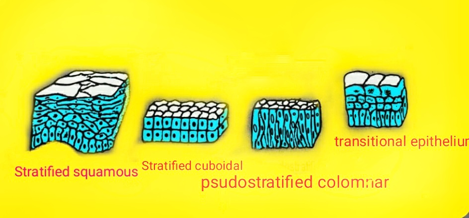
a . Stratified squamous epithelium tissue:-
- It is multi layer epithelium tissue.
It is also further classified into two types;-
1.Keratinizing epithelium tissue
2.Non keratinizing epithelium tissue.
1. Keratinizing epithelium tissue:-
- In this tissue keratin protein is present, so they are called keratinizing Tissue.
- The keratinizing tissues are found on lips, nostrils,gums,outermembrane of the tympanic membrane.
2.Non keratinizing epithelium tissue:-
- These tissue the keratin protein absent so they are called non keratinizing epithelium tissue.
- They are found at oropharynx, buccal cavity, oesophagus, inner lid of eye lids.
B. Stratified cuboidal epithelium tissue:-
- It is made up of multiple layer of cells.
- These cells are cube shaped.
- It is seen in the ducts of sweat gland
C. Stratified columnar epithelium tissue:-
- It is rare.
- It is made up of multiple layer of cells
- These cells are columnar shaped.
- It may be seen and some part of male urethra.
D. Transitional epithelium tissue:-
- It is also known as urothelium.
- It is made up of several layers of pear shaped Cells.
- It is found on urinary bladder, ureter.
*Some cells are present singly or in groups are called glands.
Classification of glands:-
a.Endocrine glands
b.Exocrine glands
c.Mixed glands.
a.Endocrine glands:-
- These glands secrete their secretions directly into the blood.
- They are ductless glands
- Ex-thyroid gland
b. Exocrine glands:-
- These glands secrete their secretions into to blood through the duct.
- Ex-sweat glands
c.Mixed glands :-
- There are some glands which having both exocrine and endocrine functions.
- Ex-pancreas gland.
2.Connective tissue:-
- The connective tissue connect various structure of our body with each other.
- It is also known as supporting tissue or communicative tissue.
- It is developed from mesoderm layer of embryo.
Components of connective tissue :-
There are are three basic components of connective tissue:-
1. Intercellularsubstance
2. Fibres of connective tissue
3. Cells of connective tissue.
1.Intercellular substances:-
- It is also known as in the matrix.
- The cells are live in intercellular substances
- The provides support to tissue.
- The help in diffusion between blood capillaries and cells
properties of intercellular substances:-
- It is a transparent.
- It is colourless and homogenous.
composition of intercellular substances:-
- the intercellular substances are composed of glycoprotein,glycosaminoglycans.
2.Fibres of the connective tissue:-
There are three main types of fibres:
A. Collagen fibers
B. Reticular fibers
C. Elastic fibers.
A. Collagen fibers:-
- Collagen fibers are found in all connective tissue.
- They are made up of polypeptides.
- They are more tough.
- They are also known as white fibers.
- These fibers are soft and flexible.
Types of collagen fibers:-
There are many five types of collagen fibers.
Type 1st:- site of occurrence is:- tendons, bones, dermis of skin.
Type 2nd:- site of occurrence is cartilage.
Type3:- site of occurrence is blood vessels, uterus, skin.
Type 4:-site of occurrence is Basal laminas
Type 5:- site of occurrence are blood vessels, fetal membrane.
B.Reticular fibers:-
- Reticular fibers are very fine fibers.
- They are arranged as net like structure.
These fibres are found in:-
- Lungs
- Lymphoid tissues.
C.Elastic fibers:-
- The elastic fibers are long, thin and highly cylindrical structure.
- These fibers are yellow in colour.
- They are also known as yellow elastic fibers.
The elastic fibres are found in:-
- Walls of major blood vessel
- Auricle(pinna)
- Epiglottis
3.Cells of the connective tissue:-
The cells of connective tissue are:-
- Mast cells
- Macrophages
- Fibroblast
- Pigment cells.
- Fat cells
- Undifferentiated mesenchymal cells
- Blood leukocytes.
1. Mast cells:-
- It is also known as ‘mastocytes’
- Mast cells are present in groups.
- These cells secrete heparin, histamine, and serotonin in allergic reactions.
Functions of the mast cells:-
- The mast cells help in wound healing.
- Defence against pathogens.
2. fat cells:-
- The fat cells are also known as lipocytes/Adipocytes
- The fat cells are large cells
- The nucleus in fatscells is flattened.
3. Macrophages:-
- The macrophages are found in highly vascular areas.
- These are irregular cells
- These cells help in phagocytosis
- They help in immunological reaction of the body
- They secrete important substances.
Ex-enzymes.
4. Fibroblast cells:-
- Fibroblast cells occur in large numbers.
- They are responsible for the production of the fibers
- Dead cells are large,flat and branching cells.
5. pigment cells:-
- Pigment cells are found in skin, Piamater and choroid part of eye.
- The main pigment of pigment cell is melanin.
- The main function of pigment cell is;-to protect the deeper tissues from ultraviolet rays.
6. Undifferentiated mesenchymal cells:-
- These cells are developed from mesoderm.
- These cells are found around the blood vessels.
- They are pleuripotant cells .
7. Blood leukocytes:-
The leukocytes are transported by blood
They are divided into two types:-
A. Lymphocytes
B. Eosinophils.
👉Classification of the connective tissue:-
1.Loose connective tissue:-
A. Adipose tissue
B. Lymphoid tissue
2. Dense connective tissue:-
A. Fibrous tissue
B. Elastic tissue
3. Cartilage tissue:-
A. Hyaline cartilage tissue
B. Fibro cartilage tissue
C. Elastic cartilage tissue
4. Bone tissue
5. Blood tissue.
1. Loose connective tissue:-
- It is found in almost every part of body.
- It contains less fibres more cells
- It contain fat cells, macrophages, and mast cell.
- These cells and fibers are loosely arranged.
Function of loose connective tissue:-
- It provides elasticity and strength.
- It connects and support the other tissue.
Types of loose connective tissue:-
1. Adipose tissue
2. Lymphoid tissue.
1. Adipose tissue:-
- It is made up of fat cells.
- It contains large fat globules.
functions of adipose tissue:-
- it occurs as a store of energy.
- It support and protect the organ.
- It help in temperature maintenance.
2.Lymphoid tissue:-
- it is also known as reticu lar tissue.
- It contain mostly reticular fibers.
- It is found in lymph nodes and all organ of lymphatic system.
2. Dense connective tissue:-
- It contains more fibers and less cells.
Types of dense connective tissue:-
There are two types of dense connective tissue……
A. Fibrous tissue
B. Elastic tissue
A.Fibrous tissue:-
- fibrous tissue contains collagen fibres with little matrix.
- It forms outer covering of bone, kidney and brain.
B. Elastic tissue:-
- Elastic tissue is made up of elastic fibres.
- They have capacity of extension and recoil.
It occurs in………
- Walls of large blood vessels.
- Trachea
- Bronchi and lungs.
3. Cartilage tissue:-
- Cartilage tissue is firmer than other connective tissue except bone.
- It consists of chondrocytes, collagen and elastic fibres.
- The mature cells of cartilage is called 'chondrocytes’
Types of cartilage tissue:-
- Hyaline cartilage
- Fibro cartilage.
- Elastic cartilage.
A. Hyaline cartilage:-
- Hyaline cartilage is smooth bluis-white issue.
- The chondrocytes are present in small groups.
- It provide flexibility and smooth surface for the movement of joints.
It founds:-
- Trachea
- Part of larynx
- Anterior and of ribs
B. Fibrocartilage:-
- Fibrocartilage is made up of collagen fibres.
- It is strongest cartilage tissue.
It is found:-……
- Intervertebral disc.
- Between articular surface of bones.
C. Elastic cartilage:-
- It is made up of elastic fibres.
- The chondrocytes lies between the fibres.
- It provides support and maintenance shape.
It is found:-
- At Wall of large blood vessels
- Epiglottis
- Panna.
I hope you like our article on Tissues and it's classification is helpful for you I suggest YouTube video that is given above for
fully understand Tissues lease it's classification watch it now and share your friends and if
you have any query related to this topic then please comment in the
comment box.
Thank you.


0 Comments
Please don't enter any spam link in comment box